
Figure 3.
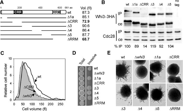
Functional analysis of the interaction between Whi3 and Cdc28. (A) Scheme depicting the Whi3 deletions described under Materials and methods. The average cell volume of exponentially growing cells containing the different deletions of Whi3 is also shown. (B) Cell extracts obtained from strains expressing the 3HA-tagged versions of Whi3 depicted in (A) were immunoprecipitated with the anti-Cdc28 antibody. The presence of Cdc28 and Whi3-3HA proteins in the corresponding immunoprecipitates (IP) and cell extracts (ce) was analyzed as described in Figure 2A. Samples obtained from an untagged strain are shown as control (no tag). Immunoprecipitation efficiencies are shown at the bottom as percentages relative to the value obtained for wild-type Whi3. (C) Volume distributions of cells carrying a wild-type WHI3 gene (wt), the whi3ΔCRR deletion that removes amino acids 121–220 (ΔCRR) or a null allele (Δwhi3). (D) Invasive growth in YEPD plates of derivative haploid Σ1278 strains carrying the 3HA-tagged versions of Whi3 depicted in (A). (E) Filamentation in SLAD plates of derivative diploid Σ1278 strains carrying the 3HA-tagged versions of Whi3 depicted in (A).