
Figure 1.
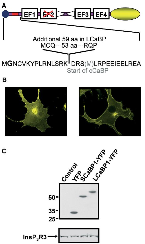
Design and expression of CaBP-YFP vectors. (A) Graphical representation of the LCaBP1-YFP and SCaBP1-YFP constructs used. The location of the additional aa in LCaBP1 compared to SCaBP1 is indicated by the red box. The central α-helix is indicated by the two triangles and the myristoylation sequence is represented by a circle. The four EF hand Ca2+-binding domains are indicated and the nonfunctional EF hand is shown by the red cross. The yellow ellipse illustrates the COOH-terminal YFP. In the lower panel, aa boundaries for both S/LCaBP and truncated cCaBP (additional methionine at the start of cCaBP shown in grey) are shown. The myristoylated glycine is in bold. (B) COS-7 cells were transiently transfected with S/LCaBP-YFP constructs. At 48 h post–transfection, cells were imaged confocally through a × 100, 1.4 n.a. oil immersion objective (Perkin-Elmer, Cambridge UK). The 488 nm line of the krypton/argon laser was used for excitation and emission collected >505 nm. (C) (Upper panel) Mobility of YFP, SCaBP1-YFP and LCaBP1-YFP was determined in transiently transfected COS-7 cells by Western blot using anti-GFP antibody. (Lower panel) Abundance of InsP3R3 was determined in cells transfected with YFP, SCaBP1, and LCaBP1 isolated by FACS.