
Figure 1.
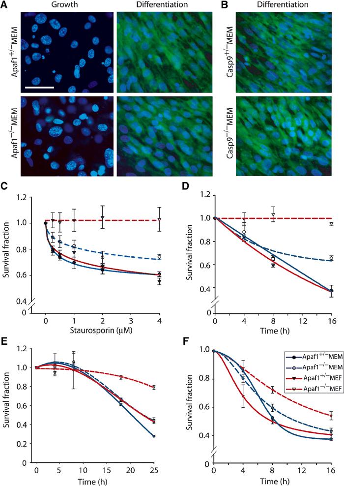
Cytotoxic drugs induce cell death in Apaf1−/− primary myoblasts but not fibroblasts. (A, B) Confocal microscopy images of Apaf1−/−, Casp9−/− and control MEMs cultured in ‘Growth' or ‘Differentiation' conditions, stained for MHC (cytoplasm—green) and counterstained with Hoechst (nucleus—blue). Bar=50 μm. (C–F) Representative MTT assays performed in duplicate on primary cultures treated with increasing concentrations of STS (C), using 2 μM STS for different duration (D); 50 μM CP (E); or 2 μM TNFα (F).