
Figure 1.
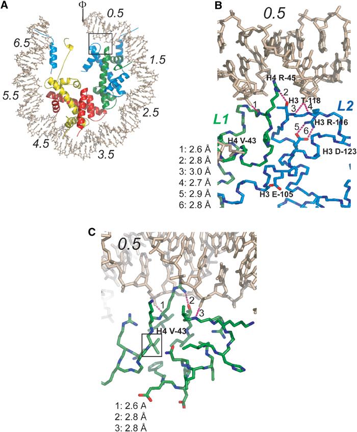
Location and structural context of histone Sin mutants. (A) Overview of the NCP structure, viewed down the superhelical axis. Only half of the DNA and associated proteins are shown. SHLs are indicated by italic numbers from 0.5 (flanking the nucleosomal dyad) to 6.5 (at the entry and exit point of the DNA). The locations of the H4 L1 loop and the H3 L2 loop are indicated in green and blue, respectively. Histone H3 is colored in blue, H4 in green, H2A in yellow, and H2B in red. The box indicates the location of Sin mutants. The nucleosomal dyad is indicated (Φ). (B) Close-up of the Sin region showing the five residues affected by Sin mutations. Dotted lines represent hydrogen bonds. (C) Location of H4 Val-43 in the hydrophobic core at the underside of the L1L2 loop arrangement. The view is similar as in (B), with a slight rotation around the y-axis.