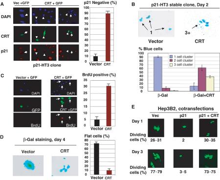
Figure 6.
Overexpression of CRT blocks the biological functions of p21. (A) CRT inhibits the translation of p21 in a stable clonal line p21-HT3. Vector +GFP (control) or CRT+GFP (ratio 10:1) were cotransfected into p21-HT3 cells, p21 was induced by IPTG and stained with specific antibodies at 16 h after IPTG addition. DAPI staining shows all cells on the field. Green shows transfected cells. Red represents p21 staining. Bar graphs show a summary of five independent experiments. (B) CRT blocks p21-mediated growth arrest. His-CRT or empty vectors were cotransfected with a β-gal plasmid, and transfected cells were visualized at day 2 by β-gal staining. Bar graphs show a summary of five independent experiments. (C) Overexpression of CRT abolishes p21-mediated inhibition of DNA synthesis. BrdU uptake was examined in p21-HT3 cells induced by IPTG, and transfected with plasmid coding for CRT or with empty vector. GFP was cotransfected to visualize transfected cells. Bar graphs show a summary of three independent experiments. (D) CRT blocks the p21-mediated formation of flat cells. CRT or empty vectors were cotransfected with plasmid coding for β-gal into p21-HT3 cells. p21 was induced by IPTG, and cells were stained for β-gal activity 4 days after p21 induction. The right shows a summary of three experiments. (E) Overexpression of CRT blocks p21 inhibitory activity in hepatoma 3B2 cells. AdTrack-p21 plasmid (coding for p21 and GFP) was cotransfected with empty vector or with His-CRT into Hep3B2 cells. Transfected cells were visualized at days 1 and 3 after transfection. The percentage of dividing green cells (two and more cells per colony) is shown below as a summary of two experiments.

