
Figure 3.
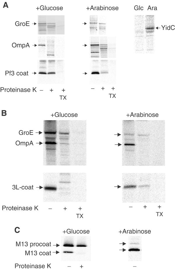
Membrane insertion of the Pf3 coat protein in vivo. Protease mapping of the Pf3 coat protein (A) and the mutant 3L-Pf3 (B) expressed in JS 7131 cells depleted of YidC (+glucose, left panels) or expressing YidC (+arabinose, right panels). Exponentially growing cells bearing the respective plasmids were induced for 10 min with 1 mM IPTG and pulse labelled with [35S]-methionine for 3 min, converted to spheroplasts and digested with 200 μg/ml proteinase K either in the absence or presence of 2% Triton X-100. The samples were immunoprecipitated with antibodies to Pf3 phage (A, B, lower panels) or to OmpA, GroEL and YidC (upper panels) and analysed by SDS–PAGE and phosphorimaging. For a control, the processing and protease accessibility of the M13 procoat protein was analysed (C).