
Figure 2.
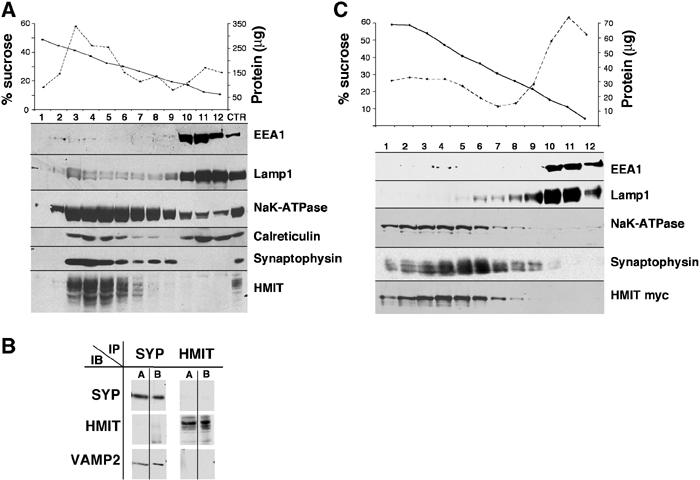
Subcellular fractionation of brain HMIT compartment. (A) Fractionation of mouse brain membrane preparation on a sucrose density gradient (10–50%). Fractions were analyzed for protein and sucrose concentration (top). HMIT and several membrane fraction markers were detected by Western blot. (B) Immunoprecipitation of HMIT- and synaptophysin-containing vesicles. Magnetic beads coupled with antibodies against HMIT or synaptophysin were incubated in the presence of mouse brain membrane proteins. The immunoprecipitated vesicles were first solubilized with Triton X-100 and the proteins remaining attached to the beads were removed by an SDS-containing buffer. Both the Triton X-100-solubilized proteins (lanes A) and the SDS-elution proteins (lanes B) were analyzed by Western blot for the detection of HMIT, synaptophysin or Vamp2. (C) Fractionation on sucrose density gradient of homogenates of HMIT-myc-expressing PC12, as described in (A).