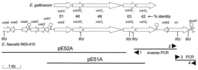
FIG. 1.
Genetic organization of the vanE locus from E. faecalis N00-410 showing regions cloned into plasmids and regions isolated by inverse PCR and PCR. The direction of transcription is indicated by arrows, and putative stem-loop structures are indicated. Primers used in inverse PCR and PCR are indicated by black arrowheads: primer 1 is vanRE-1, primer 2 is vanSE-DN1, primer 3 is Eint-DN1, and primer 4 is gua-DN1. The organization of the vanC1 locus from E. gallinarum is shown at the top. Percent identities of the corresponding vanC1 and vanE operon proteins are shown. RV indicates EcoRV sites.

