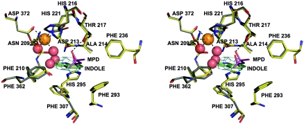
FIG. 3.
Stereo view of the active site of NDO-R. MPD from the buffer is found bound in the active site of the native enzyme (structure 1), and indole is bound in the substrate-soaked crystal structure (structure 2). Structure 1 is in gray and structure 2 in yellow. Four water molecules found in the cavity of the active site in structure 1 are shown as red spheres. Two of these waters form part of the octahedral coordination of the iron (dashed lines). A hydrogen bond between MPD and T217 (dashed line) suggests that polar groups on the nonhydroxylating ring of the substrates may interact with T217. A 2FoFc omit map for indole contoured at 0.8 sigma is shown in cyan.

