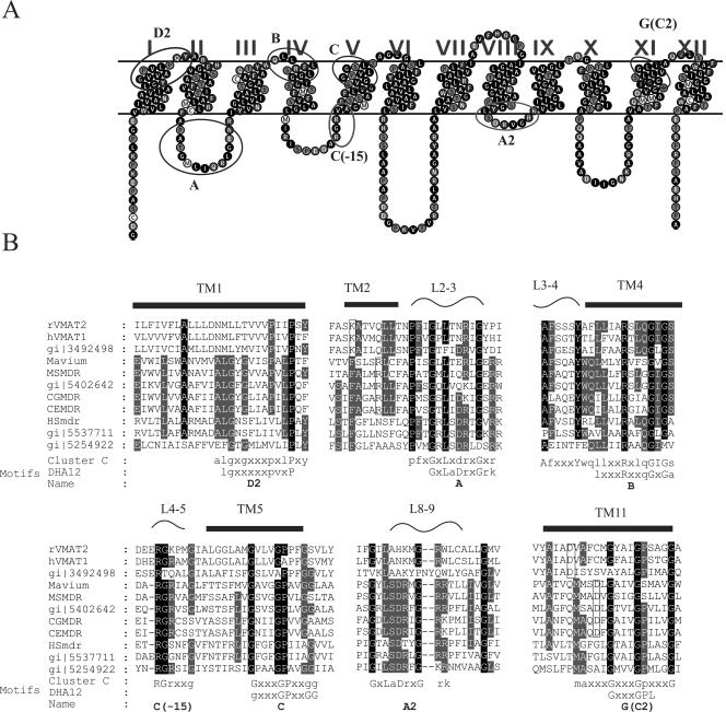
FIG. 2.
Sequence analysis of cluster C. A. A representative topology suggested for DHA12 proteins contains 12 transmembrane domains divided into two halves by a long cytoplasmic loop between TM6 and TM7. The topology shown is that of MSmdr. Motifs from the DHA12 family are marked with black circles and motifs specific for cluster C are marked with gray circles. B. Conserved regions in the sequence alignment of cluster C. At the bottom of the alignment are consensus sequences in which capital letters represent frequency occurrence greater than 90% (dark shading) and small capitals represent frequency occurrence greater than 50% (light gray shading). The consensuses are compared with the DHA12 motifs defined by Paulsen et al. (27). The arrows above the sequences point to residues involved in putative ion pairs (open rectangles).

