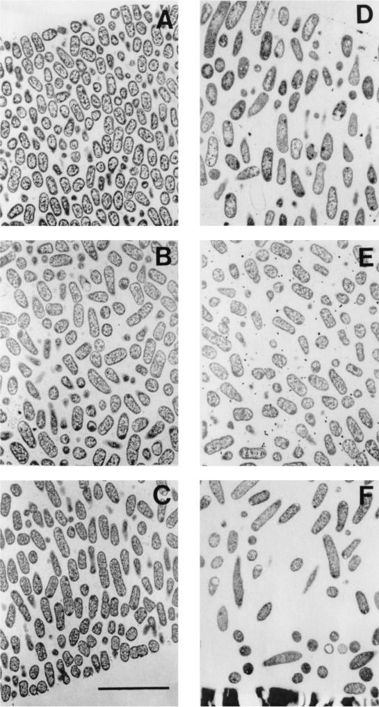
FIG. 1.
Transmission electron micrographs of β-lactamase-positive K. pneumoniae colony biofilms from the untreated 12-h control (A to C) and exposed to 1.8 μg of ciprofloxacin per ml for 12 h (D to F). (A and D) Spots near the air interface, which is just visible in the upper left (A) and right (D) corners; (B and E) locations near the middle of the biofilm; (C and F) regions near the membrane. The membrane is visible in the bottom of panel F. The membrane detached from the specimen shown in panel C; the former location of the membrane was along the bottom right-hand corner of the panel. Scale bar, 5 μm.

