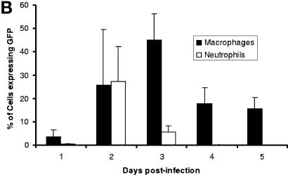
FIG. 3.
Dissemination of Y. pestis(pACGFP) into cells of the innate immune system following subcutaneous infection of BALB/c mice. The spleens from groups of five BALB/c mice were removed on days 1, 2, 3, 4, and 5 postinfection. Neutrophils (Ly-6G+ cells) and macrophages (CD11b+ cells) were isolated using Minimacs beads, stained for the presence of Ly-6G and CD11b, fixed in 4% paraformaldehyde for 24 h at 4°C, and analyzed by flow cytometry. (A) Dot plots from CD11b+ and Ly-6G+ populations on each day postinfection, indicating the presence of GFP fluorescence. Regions from which CD11b+ and Ly-6G+ cell data were gated are also indicated. (B) Percentages of CD11b+ (solid bars) and Ly-6G+ (open bars) cells expressing GFP over the course of infection. The data are means with 95% confidence limits.


