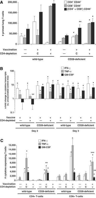
FIG. 1.
T-cell priming and production of IFN-γ, TNF-α, and GM-CSF transcript and protein by lung T cells after B. dermatitidis infection in vaccinated and unvaccinated wild-type and CD28−/− mice. (Top panel) Priming of lung T cells as measured by surface expression of CD44 on cells analyzed 4 days postinfection. The data are an average of six to nine mice/group. Mice were infected with 2 × 10³ yeast. *, P < 0.05; **, P < 0.1 (versus vaccinated wild-type controls). Controls (C) represent vaccinated mice treated with rat IgG. (Middle panel) n-fold changes in lung cytokine transcripts in vaccinated wild-type and CD28−/− mice versus unvaccinated controls as measured by real-time PCR. The data represent an average of three to five mice/group. Mice were infected with 2 × 10³ yeast and analyzed 2 and 3 days after infection. *, P < 0.05 (versus unvaccinated controls). Cytokine transcripts did not differ significantly in vaccinated CD28−/− mice versus wild-type controls, except for a marginal difference in GM-CSF transcript at day 2. **, P = 0.06. (Bottom panel) Intracellular cytokine production by CD44+ CD4+ and CD44+ CD8+ cells. The data represent an average of six to nine mice per group. Mice were infected with 2 × 10³ yeast and analyzed 4 days after infection. *, P < 0.008 (versus corresponding unvaccinated mice); **, P ≤ 0.02; ***, P < 0.07 (versus corresponding vaccinated wild-type controls). Controls (C) represent vaccinated mice treated with rat IgG.

