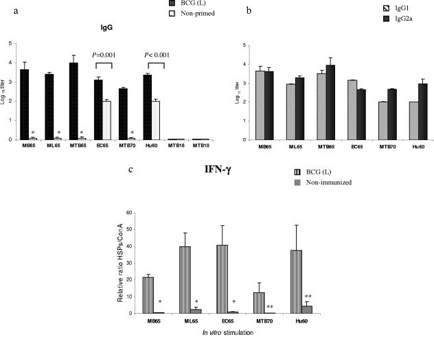
FIG. 3.
(a) HSP-specific IgG responses induced in BALB/c mice primed with live (L) BCG and boosted with various HSPs once. Control mice received only HSPs once. Serum samples were obtained from the mice 7 days after the protein immunization. The presence of HSP-specific antibodies was detected by ELISA using serial dilutions of individual sera from three mice. Significant differences between groups following a Student's t test are depicted with asterisks (*, P < 0.005 with the respective control group). (b) HSP-specific IgG1/IgG2a profile. Titers are expressed as the log10 of the reciprocal of the highest dilution at an OD of 1.0. Background reactivity of the sera with BSA at the corresponding dilution was subtracted. (c) In vitro IFN-γ production by the splenocytes stimulated with various HSPs from mice primed with live BCG or not primed. Splenocytes of individual mice from each group were stimulated separately in triplicate with 10 μg/ml of each HSP and with 2 μg/ml of ConA (positive control) in 96-well cell culture plates and incubated for 72 h at 37°C in 5% CO2. Cell supernatants were collected, pooled, and assayed for the presence of IFN-γ by ELISA. Since the absolute values varied from experiment to experiment, to facilitate comparisons we normalized all values in relation to ConA stimulation. Data are expressed as the ratio between HSP and ConA stimulation multiplied by 100. Values represent means of triplicate samples with SD. *, P = 0.05; **, P < 0.01 with the respective controls.

