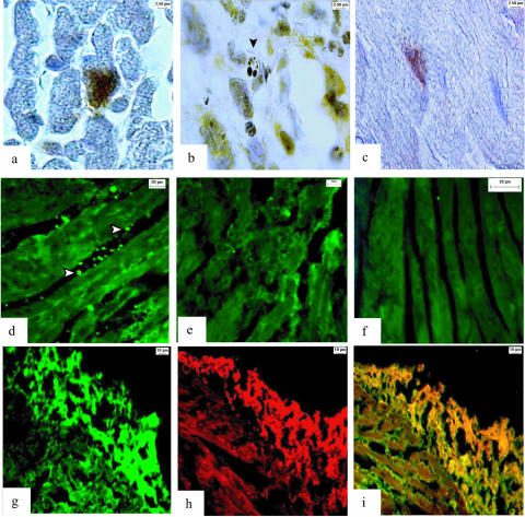
FIG. 2.
Cardiac apoptosis in relapsing fever and Lyme borreliosis. a) Cardiomyocyte with positive TUNEL staining in the nucleus (brown color) of one Igh6−/− mouse persistently infected with B. turicatae serotype 2. Magnification, 1,000×. b) Apoptotic body (arrow) and expression of Bax (yellow color) in the heart of a scid mouse infected with B. turicatae. 1,000× magnification. c) TUNEL staining in the nucleus of a cardiomyocyte from an immunosuppressed nonhuman primate persistently infected with B. burgdorferi. 1,000× magnification. d) Anti-caspase-1 antibody shows green fluorescence in mononuclear cells from an Igh6−/− mouse infected with B. turicatae serotype 2. 400× magnification. e) Negative control section shows lack of staining in an area of severe inflammation and cardiac fiber degradation incubated with 2 μg/ml of rat IgG as a negative control. 400× magnification. f) Myocardium from an uninfected Igh6−/− mouse stained with anti-caspase-1 antibody shows no signal. 400× magnification. g) Periphery of the left ventricle near the atrium from an Igh6−/− mouse infected with serotype 1 and FITC stained with anticaspase-1 antibody (200× magnification). h) Consecutive section from the same mouse shows red fluorescence staining (TRITC) after incubation with anti-F4/80 antibody, a marker of activated macrophages. i) Double immunofluorescence staining with anti-caspase-1 (FITC) and anti-F4/80 (TRITC) antibodies shows yellow color, indicating colocalization, in the pericardium and membranes from nearby cardiomyocytes.

