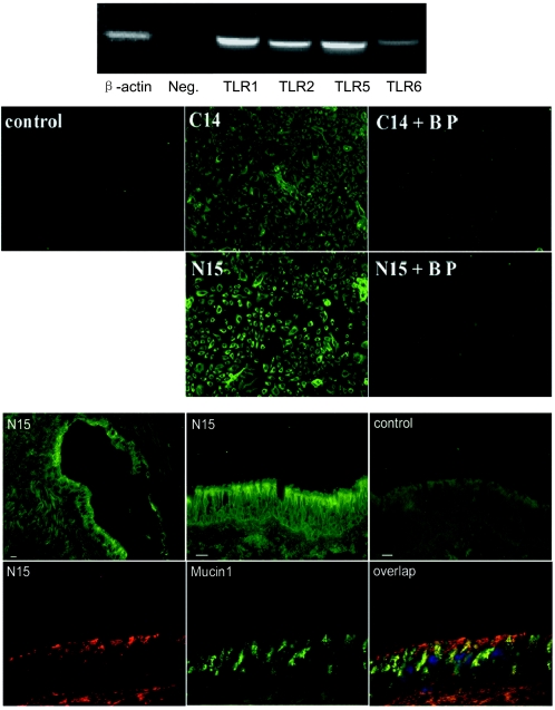
FIG.3.
TLR5 is expressed in HAECs and on the apical surface of human airway epithelium. (Upper panel) RT-PCR using TLR-specific primers was performed to test the expression of TLRs in total mRNA of HAECs. The samples were a human β-actin positive control (587 bp), a negative control (Neg.) in which primers were omitted from the reaction mixture, human TLR1 (615 bp), human TLR2 (612 bp), human TLR5 (600 bp), and human TLR6 (625 bp). (Middle panel) Expression of TLR5 in HAECs. HAECs cultured on glass slides were stained with human TLR5 antibodies generated against either the TLR5 C terminus (C14) or the TLR5 N terminus (N15). Specific blocking peptide (BP) of C14 or N15 was used to neutralize the staining, as described in Materials and Methods. The control shows staining after replacement of the primary antibody by an isotype-matched IgG. (Lower panel) Expression of TLR5 in airway epithelium. Cryosections of human trachea were blocked with 10% goat serum and stained with primary human TLR5 antibody (N15) and then FITC-labeled secondary antibodies. Images stained with N15 were obtained at low amplification (top left image) or high amplification (top middle image). The control (top right image) shows staining after antibodies were replaced by an isotype-matched IgG. Cryosections of human trachea were also stained with TLR5 N15 antibody (recognized by TRITC-labeled secondary antibody) (bottom left image), human Mucin1 antibody (recognized by FITC-labeled secondary antibody) (bottom middle image), and 4′,6′-diamidino-2-phenylindole (DAPI) at the same time. Sections were observed using a triple filter (bottom left image). Bars = 15 μm.

