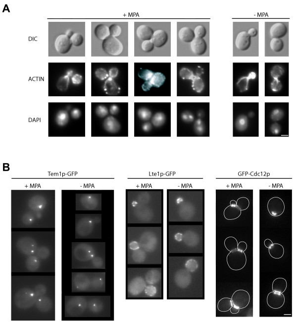
Figure 5.
Localization of several cellular structures or fusion proteins in cells with two daughters. A. Cells were untreated (right panel) or treated (left panel) with 100 μg/mL MPA for 4 hours. DIC (top lane), actin Alexa-phalloidin staining (middle lane) and DAPI staining (bottom lane) are shown. B. Localization of endogenous Tem1p-GFP (left panel), endogenous Lte1p-GFP (middle panel) and GFP-Cdc12p expressed from a centromeric plasmid under the control of its own promoter (right panel) in cells untreated (right of each panel) or treated for 4 hours with 100 μg/mL MPA (left of each panel). Only cells displaying two buds are shown in the case of MPA treatment. Bar: 2 μm.

