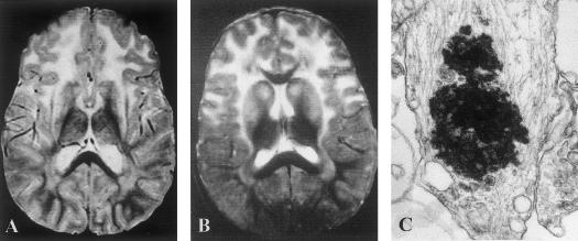
Figure 1.
A and B, Typical T2-weighted MRI images of brains of patients with infantile Alexander disease. A, Classical form (patient 7). B, Mild form (patient 3). Note that both patients show the high signal intensity of white matter, predominately in the frontal area, and of the basal ganglia. C, Rosenthal fiber in Alexander disease viewed by electron microscopy (patient 15); original magnification ×28,500.

