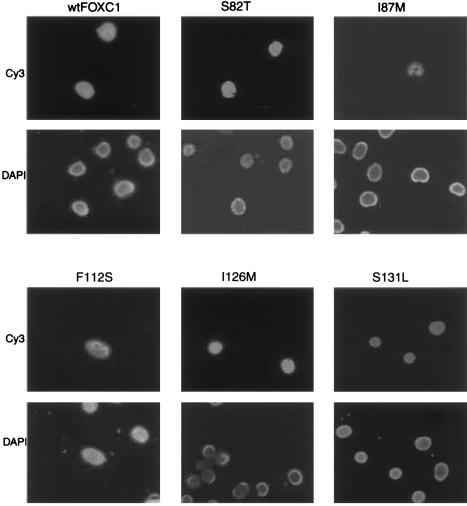
Figure 4.
Immunofluorescence of Xpress epitope–tagged FOXC1 proteins. COS-7 cells were transiently transfected with the FOXC1 pcDNA4 His/Max constructs. In the upper six panels, the Xpress epitope–tagged recombinant FOXC1 proteins are localized to the nucleus, indicated by Cy3 fluorescence. The lower six panels indicate the position of the nuclei, by staining with DAPI. Note that the Cy3 fluorescence in the I87M transfection is weaker than it is in the other mutations.

