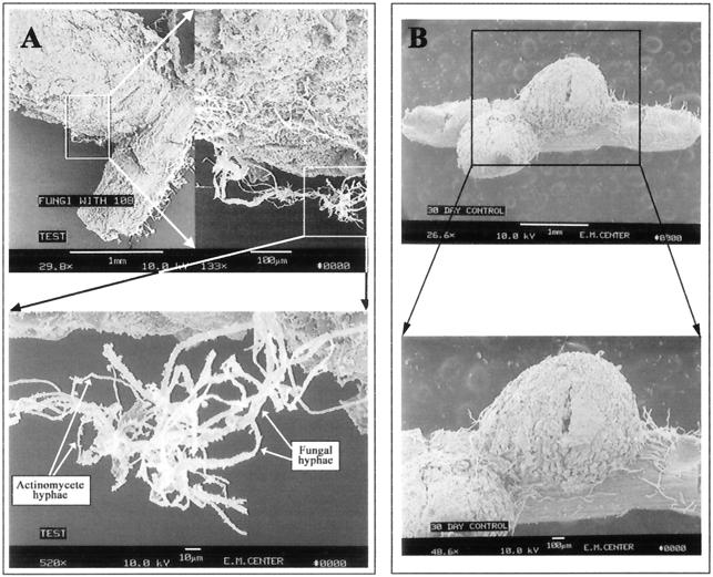
FIG. 7.
SEM micrographs of the root nodule surface. (A) Thirty-day-old colonized nodule, with intense surface colonization by actinomycete hyphae (top panel; bar = 1 mm) and fungal hyphae interlocked with actinomycete hyphae on the colonized root surface (bottom panel; bar = 10 μm). (B) Thirty-day-old control nodule, with absence of colonization by any actinomycete; the filaments are root hairs (bar = 1 mm [top panel] and 100 μm [bottom panel]).

