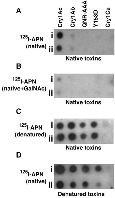
FIG. 3.
Reverse-dot blot analyses of purified toxins. Five micrograms (row i) and 1 μg (row ii) of purified toxins were dot blotted as described in the legend to Fig. 2. (A) Toxin on membrane exposed to 125I-APN. (B) Toxin on membrane exposed to 125I-APN plus 100 mM GalNAc. (C) Toxin on membrane exposed to denatured 125I-APN. 125I-APN was denatured as described in Materials and Methods. (D) Purified toxins were denatured as described in Materials and Methods before dot blotting onto the membrane and exposure to 125I-APN. All preparations were incubated for 1 h, washed, and autoradiographed.

