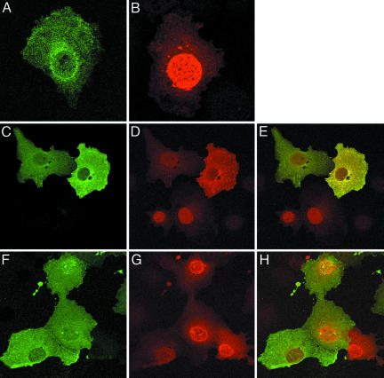
Fig. 4.
Intracellular localization of Tat and HIC. (A) Cos7 cells expressing Tat. (B) Cos7 cells expressing HIC. (C-E) Colocalization of HIC and Tat. (C) HIC (green) remains localized in the cytoplasm. (D) Redistribution of Tat (red) in the cytoplasm. (E) Merged images showing colocalization of HIC and Tat (yellow). (F-H) The I-mfa domain is involved in the sequestration of Tat. When HIC(2-144) and Tat are coexpressed, HIC localizes in the cytoplasm (F) and Tat localizes in the nucleus (G). Merged images (H) confirmed the absence of colocalization. Arrows indicate cells expressing Tat only.

