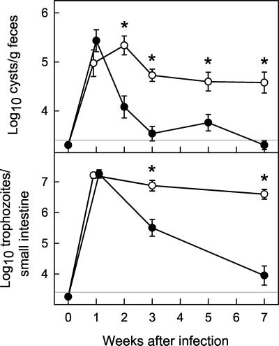
FIG. 1.
Primary G. muris infections of B-cell KO mice. B-cell KO mice (○) and wild-type littermate C57 control mice (•) were infected orally with 104 G. muris cysts. Infection intensity was assessed at the indicated times after infection by determining stool cyst output (top panel) and total trophozoite numbers in the small intestine (bottom panel). All data are means ± SEM from nine or more mice for each data point. The detection limits of the assays are indicated by the gray line in each panel. Asterisks indicate values of B-cell KO mice that are significantly different from controls at the same time point (P < 0.01 by rank sum test).

