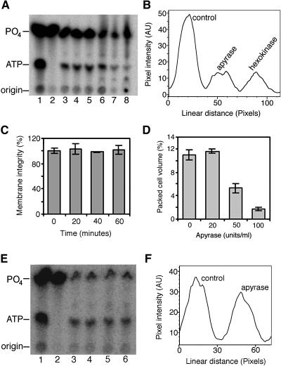
Figure 1.
Extracellular ATP and Cell Viability of Arabidopsis Cell Cultures.
(A) ATP and phosphate (PO4) standards (lane 1) and [32P]H3PO4 fed to three independent cell cultures and medium aliquots taken at time 0 (lane 2) and 1 h later (lanes 3 to 5) showing the de novo synthesis and secretion of ATP. At 1 h, solutions of ATP traps were added, and 6 h later, medium samples were taken from the control culture (lane 6) and from cultures treated with 100 units/mL apyrase (lane 7) and glucose–hexokinase (lane 8).
(B) Line graph of a pixel intensity profile showing relative levels of ATP in the 6-h samples (lanes 6 to 8) in (A).
(C) Plasma membrane integrity, measured by Evans blue staining, of cell cultures in (A) over the first 1 h.
(D) Dose response of cell viability to treatment with apyrase.
(E) ATP and phosphate standards (lane 1), [32P]H3PO4 (lane 2), and medium aliquots 1 h after adding radioactive phosphate (lanes 3 and 4) and just before adding 20 units/mL apyrase. Medium from control cells (lane 5) and cells treated with 20 units/mL apyrase (lane 6) 6 h after enzyme treatment.
(F) Line graph of a pixel intensity profile across the ATP bands in lanes 5 and 6 of (E).
Data and error bars represent means ± sd (n = 3). AU, arbitrary units.

