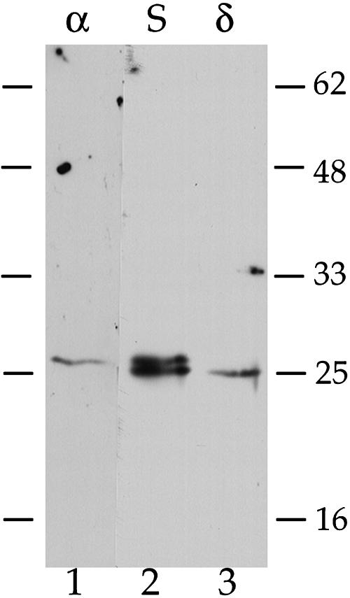
Figure 10.
Immunoblot Analyses of TIPs in Pea Seed Membrane Proteins.
Protein extracts from dry, mature pea seeds were prepared as described (Jauh et al., 1999). Three 50-μg samples were electrophoresed through a 15% acrylamide SDS-PAGE gel, where molecular mass markers bracketed either side. After transfer to nitrocellulose, the membrane was stained with Ponceau red to identify the lanes. Three strips were excised, where notches at the bottom were placed to allow exact alignment of the strips: strip 1 (α), markers + pea proteins, incubated with isoform-specific anti-α-TIP peptide antibodies (Jauh, et al., 1999), 1 μg/mL; strip 2 (S), pea proteins, incubated with anti-α-TIP antiserum (Johnson et al., 1989), 1:2000; strip 3 (δ), pea proteins plus markers, incubated with isoform-specific anti-δ-TIP peptide antibodies, 10 μg/mL. After overnight incubation, the strips were processed for detection by chemiluminescence. The exposures presented are 10 s for strip 1 and 2 s for strips 2 and 3. Bars to either side indicate positions of molecular mass markers in kilodaltons.

