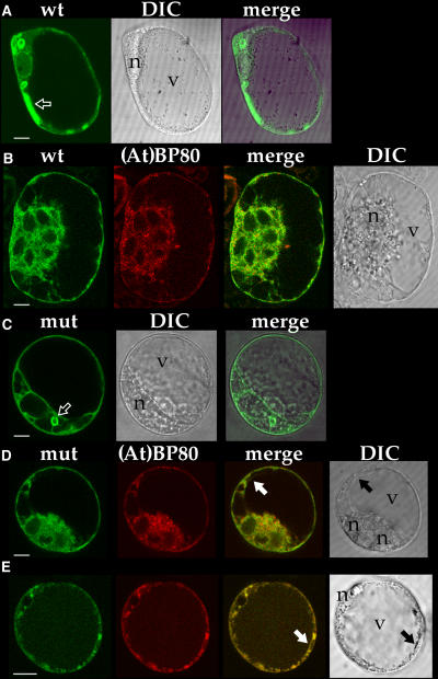
Figure 4.
Expression of GFP-Tagged α-TIP and mRFP-Tagged Proaleurain-FLAG-(At)BP80b Transmembrane Domain and Cytoplasmic Tail [(At)BP80b-mRFP] Constructs in Protoplasts.
Cells were studied 16 to 20 h after electroporation. The images in line under each letter came from one cell; green indicates GFP, red indicates mRFP, merge indicates an image resulting from superimposition of the two images immediately to the left, and DIC (differential interference contrast) indicates a transmitted light image from that specific cell.
(A) Cell expressing GFPαTIP (wt); arrow indicates GFP-tagged structure that did not have a visible lumen when sequential optical sections were collected at 0.5-μm steps.
(B) Cell expressing GFPαTIP (wt) and (At)BP80b-mRFP. This cell has six nuclei, one of which is indicated with an “n.”
(C) Cell expressing GFPαTIPΔ (mut, green); arrow indicates brightly labeled vacuole.
(D) and (E) Cells expressing GFPαTIPΔ (mut, green) and (At)BP80b-mRFP (red); arrows indicate central vacuole tonoplast tagged with GFP. n, position of nucleus; v, central vacuole.

