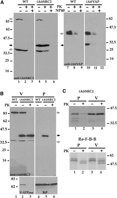
Figure 8.
Proteinase K Protection Assay.
Nontransformed (WT) and (At)SRC2- or (At)VAP-transformed protoplasts were separated into vacuole (V) and pellet (P) fractions.
(A) Vacuoles were digested (+) or not digested (−) with proteinase K (PK) in the presence (+) or absence (−) of Nonidet P-40 (NP40) detergent. Proteins were detected on immunoblots using anti-(At)SRC2 or anti-(At)VAP recombinant protein antibodies.
(B) Vacuole and pellet fractions were separately treated (+) or not (−) with proteinase K and blotted for the detection of (At)SRC2 (top panel), subunit A of the peripheral complex of V-ATPase (bottom panel to the left), and BiP (bottom panel to the right). Closed and open arrows show the positions of (At)SRC2 or (At)VAP and cross-reacting endogenous proteins, respectively. Positions of molecular mass standards are indicated in kilodaltons at the side.
(C) Protoplasts transfected to express (At)SRC2 (top) or Re-F-B-B (bottom) were labeled with [35S]Met + Cys for 1 h and then fractionated into pellet and vacuole fractions. Aliquots were separately treated (+) or not (−) with proteinase K, immunoprecipitated with anti-(At)SRC2 recombinant protein antibodies (top) or with anti-aleurain antibodies (bottom), and then separated by SDS-PAGE and visualized by fluorography.

