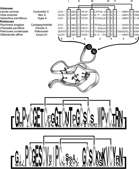
Figure 1.
Representative Sequences of Cyclotides from the Violaceae and Rubiaceae.
All peptides are head-to-tail cyclized, and the sequences are written starting from the presumed N-terminal cleavage point in the linear precursors (Jennings et al., 2001; Dutton et al., 2004). Sequences are aligned based on the conserved Cys residues, which are numbered in Roman numerals according to the order in which they appear in the linear precursor. Loops corresponding to the backbone segments between Cys residues are numbered for reference. Loop 6 is formed at the cyclization point of the linear precursor. References for the listed cyclotides are as follows: cycloviolin C (Hallock et al., 2000), varv A (Claeson et al., 1998), Hypa A (Broussalis et al., 2001), cyclopsychotride (Witherup et al., 1994), circulin A (Derua et al., 1996), palicourein (Bokesch et al., 2001), and kalata B1 (Saether et al., 1995). A ribbon diagram of the cyclotide framework based on the structure of kalata B1 (PDB ID 1NB1) with the cystine knot core rendered and numbering of loops is shown in the middle of the figure. At the bottom of the figure are two sequence logos (Crooks et al., 2004) to graphically illustrate sequence conservation within known cyclotides. The size of the one-letter amino acid code reflects the relative abundance of that residue in known cyclotides. Separate logos are shown for Möbius (top) and bracelet (bottom) subfamilies of cyclotides (Craik et al., 1999).

