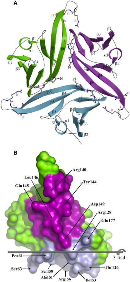
Figure 5.
The Crystallographic Trimer of ToxA.
(A) A ribbon diagram of the ToxA trimer as seen in the asymmetric unit of the form-II structure looking down the threefold axis is shown. The three chains are labeled and shown in green, blue, and purple. The four N-terminal residues and the RGD residues are shown as stick models.
(B) The two trimerization surfaces of ToxA. For this image, the green molecule from (A) is separated from the others and rotated 90° around the vertical. The narrow spine-like interface interacting with the purple molecule is facing the reader. The broad vise-shaped interface interacting with the blue molecule faces down. For trimer assembly, the blue surface of one molecule meshes with the purple surface of another. Residues having ≥40-Å2 surface area buried at the interface are labeled. The threefold axis is indicated and labeled. This figure was generated using Pymol (www.pymol.org).

