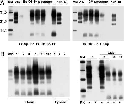
Fig. 1.
Western blot analysis of PrPres in the brain or spleen of tg338 mice inoculated with brain material from Nor98 sheep (A) or discordant sheep and goat (B). Tissues collected from terminally diseased mice were homogenized, PK-treated, and analyzed individually. (A) Transmission of Nor98 (Lindås isolate; see Table 1). Data obtained with brain (Br) or spleen (Sp) from several primary (Left) or secondary (Right) infected mice are presented. PrPres, detected only in the brain, shows a profile that is unique compared with that seen in mice of this same line inoculated with natural scrapie isolates (designated 21K or 19K). NI, noninfected brain control. (B) Primary transmission of discordant isolates. The isolates are identified by a number as in Table 2 and include one from goat (7) and three from ARR/ARR sheep (Right, 8-10). Typical data obtained with tg338 mouse brain or spleen material are shown. (Left) The PrPres profiles observed are all similar to those produced by the Nor98 agent (Rauland: Nor). (Right) PK treatment (+) of healthy (NI) or discordant (9) ARR/ARR sheep-brain material leads to disappearance and shifting of the PrP signal, respectively, whereas PrP signals of similar intensities are seen with PK-untreated (-) samples. MM, molecular markers.

