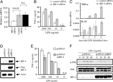
Fig. 4.
The levels of IRF-4 expression in macrophages affect the production of TNF-α. (A) Peritoneal macrophages were transfected with IRF-4-specific siRNA, control siRNA, or Lipofectamine 2000 only. Eight hours later, the expression of IRF-4 mRNA was determined by real-time RT-PCR. (B) Peritoneal macrophages were transfected with control siRNA (open bars) or IRF-4 specific siRNA (filled bars), cultured for 5 h, washed, and cultured in the presence of LPS for an additional 24 h. (C) Peritoneal macrophages transfected with control or IRF-4-specific siRNA were cultured with LPS (1 μg/ml) for the indicated period. The expression levels of TNF-α and IL-12 mRNA were determined by real-time RT-PCR analysis. (D) RAW264.7 cells were transfected with pcDNA3 or pcDNA3-mIRF-4. After5hof culture, cell lysate was separated by 12.5% SDS/PAGE. The blot was probed with anti-IRF-4 Ab, stripped, and reprobed with anti-Flag mAb. The same sample was separated by SDS/PAGE, blotted, and probed with anti-actin Ab. (E) RAW264.7 cells were transfected with pcDNA3 or pcDNA3-mIRF-4, cultured for 5 h, and stimulated with LPS for 8 h. (F) RAW-264.7 cells transfected with empty vector (pcDNA3) or with IRF-4 (pcDNA3-mIRF4) were stimulated with LPS. Cell lysate was separated by 12.5% SDS/PAGE. The blot was probed with anti-phospho-JNK Ab, stripped, and reprobed with anti-JNK Ab. The same blot was stripped and reprobed with anti-IRF-4 Ab.

