
Figure 2.
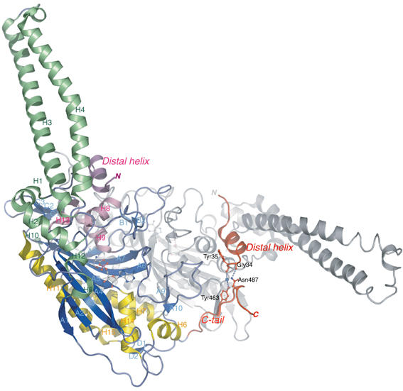
Ribbon representation of the three-dimensional crystal structure of B. taurus mt SerRS at 1.65 Å resolution, depicted in different colors for three α-helical bundles and β-sheet strands. The mitochondria-unique N-terminal distal helix (of monomer 2) and C-tail (of monomer 1) are colored in red, with the stick representations showing the mutual interaction sites. The second monomer is drawn as gray ribbons for clarity. Secondary structure elements are denominated corresponding to bacterial SerRS (Cusack et al, 1990). This figure and subsequent figures were composed using PyMol (http://pymol.sourceforge.net/).