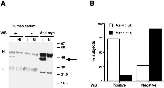
FIG. 2.
(A) Western blots (WB) with human positive (+) and negative (−) sera as well as with anti-Myc MAb of anti-Myc immunoprecipitates from lysates of noninduced (NI) and IPTG-induced (I) E. coli transformants expressing the Mce2-Myc fusion protein. Heavy (H) and light (L) chains of the immunoprecipitating anti-Myc MAb as well as the Mce2-Myc protein (arrow) are indicated. Numbers on the right are molecular sizes in kilodaltons. (B) Relative distributions of M. tuberculosis-positive (black bars) and -negative (white bars) subjects with respect to IgG reactivity to Mce2-Myc protein in immunoblot.

