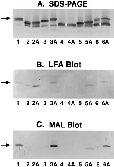
FIG. 3.
Sialylation of LOS in N. meningitidis grown in TSB with and without CMP-NeuNAc. (A) Silver-stained LOSs on SDS-PAGE; (B) LFA blot for detecting sialylated LOSs; (C) MAL blot for detecting α2,3-linked sialic acid. Lanes 1, M986 LOS markers as in Fig. 2; lanes 2 and 2A, 126E (C:L1) strain grown in the absence and presence of 200 μg of CMP-NeuNAc/ml, respectively; lanes 3 and 3A, strain M986-NCV (L3,7); lanes 4 and 4A, strain A1 (A:L8); lanes 5 and 5A, strain 7880 (A:L10); lanes 6 and 6A, strain M978 (B:L8). The arrows and asterisks are as described in the legend of Fig. 2.

