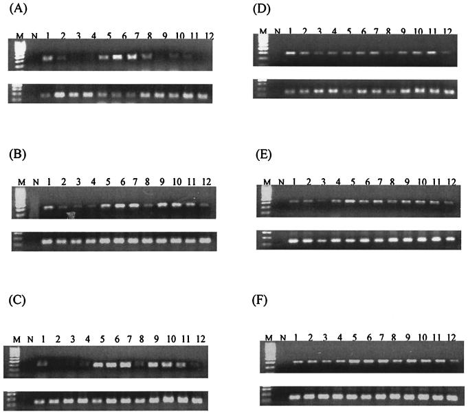
FIG. 2.
Expression of IFN-γ mRNA in the lungs (A and D), livers (B and E), and spleens (C and F) of BALB/c (A, B, and C) and C57BL/6 (D, E, and F) mice as determined by RT-PCR. In each set of gels, the top panel depicts IFN-γ and the bottom panel depicts the corresponding GAPDH. Lanes M, 100-bp molecular size marker; lanes N, no-template control; lanes 1 to 3, three infected mice at 24 h postinfection; lanes 4, control mock-infected mouse at 24 h postinfection; lanes 5 to 7, three infected mice at 48 h postinfection; lanes 8, control mock-infected mouse at 48 h postinfection; lanes 9 to 11, three infected mice at 72 h postinfection; lanes 12, control mock-infected mouse at 72 h postinfection.

