
Figure 4.
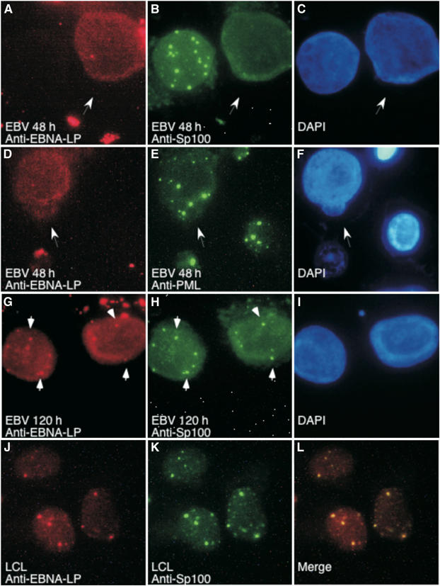
Sp100, but not PML, is displaced from PML NBs following EBV infection of B lymphocytes. At 48 h after EBV infection, Sp100 (green, B) was displaced from PML NBs in nearly all infected cells. EBV-infected cells were identified by staining with anti-EBNA-LP antibodies (red, A, D, G, J). PML (green, E) was not displaced from NBs during EBV infection. At 120 h after infection, both Sp100 (green, H) and EBNA-LP localized to PML NBs. In LCLs, both Sp100 (green, K) and EBNA-LP (red, J) localized to NBs. Overlap between Sp100 and EBNA-LP appears yellow in (L). White arrows in (A–F) indicate EBV-infected cells. White arrows in (G, H) point to examples of EBNA-LP and Sp100 in NBs. DAPI staining indicates the location of nuclei in (C, F, I).