
Figure 5.
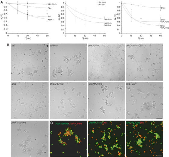
APP family proteins are required for cell adhesion. (A) Quantitative cellular aggregation of WT, APP knockout (APP−/−), APLP2 knockout (APLP2−/−), or APP/APLP2 double-knockout MEF cells (Dko). MEF cells were aggregated in suspension under calcium- and magnesium-free conditions for 1 h at 80 r.p.m. The number of particles (single cells and cell clusters) was counted at the indicated time points (Nt). The relative decrease in particle counts compared to t=0 (Nt/N0) is shown over time as a measure of aggregation of the different MEF cells (n⩾3, ±s.d., t-test). APP−/− MEFs were compared with APP retransfected cells (APP−/−APPre), and APLP1 (DkoAPLP1re) or APLP2 (DkoAPLP2re) rescued Dko cells were compared with parental Dko cells. (B) Typical micrographs of the different MEF lines after 60 min of aggregation. APLP2−/− and Dko cells were additionally aggregated in the presence of 1 mM Ca2+ to induce cadherin-mediated adhesion (scale bar=100 μm). (C) Equal amounts of differentially labeled Dko, DkoAPLP1re, and DkoAPLP12re cells (as indicated) were coaggregated for 60 min and analyzed by fluorescence microscopy (scale bar=100 μm).