
Figure 1.
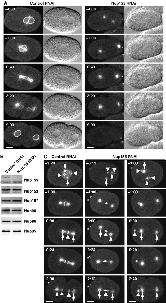
Depletion of Nup155 from C. elegans embryos affects nuclear appearance. (A) Embryos expressing GFP-β-tubulin and YFP-lamin from control (left panels) or Nup155-depleted worms (right panels) were observed by time-lapse microscopy. In this and other figures, images are shown with indication of time (minutes:seconds) relative to anaphase onset and with embryos anterior to the left. In the Nup155 RNAi embryo, neither pronuclei nor nuclei were visible. See Supplementary video 4. (B) Western blot analysis of embryonic lysates from mock-depleted (left lane) or Nup155-depleted worms (right lane) with antisera against various nucleoporins. The asterisk marks a protein that crossreacts with the Nup155 antibody, and demonstrates equal gel loading. (C) Control (left) or Nup155-depleted (middle and right) embryos expressing GFP-β-tubulin and GFP-histone H2B were observed by time-lapse microscopy. In selected panels, centrosomes are indicated with arrows whereas triangles point to chromatin. See Supplementary video 5. Scale bars, 10 μm.