
Figure 7.
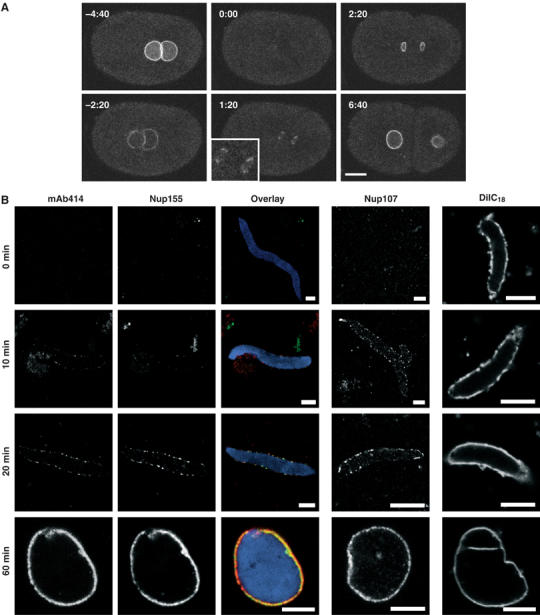
Dynamics of Nup155. (A) Still images from time-lapse microscopy recording of C. elegans embryo expressing GFP-Nup155. Images show progression from pronuclear meeting in the one-cell embryo to the two-cell stage. The inset shows a magnification of initial GFP-Nup155 recruitment to the two reforming nuclei during anaphase (1:20). See Supplementary video 8. Scale bar, 10 μm. (B) NE assembly reactions were fixed in 4% paraformaldehyde at the time points indicated. The three columns on the left, which show immunodetection of Nup155 (green), mAb414 (red) and DAPI (blue), are identical samples and merged in the overlaid images. Parallel reactions for Nup107 immunodetection or that were stained with the membrane dye DiIC18 before fixation are on the right. Scale bars, 5 μm.