
Figure 4.
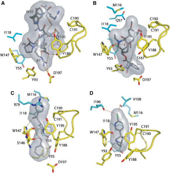
Expanded views of the bound ligands. Hydrogen bonding of key residues for the bound (A) ImI, (B) MLA, (C) LOB, and (D) EPI, viewed from inside of the ion channel vestibule looking in a radial direction. Ligands are bound between the Cys190–Cys191 disulfide on the left and Trp148 on the right. Labels for the ImI residues are italicized. The molecular surfaces of the ligands are in gray. Subunit coloring is identical to that in Figure 3.