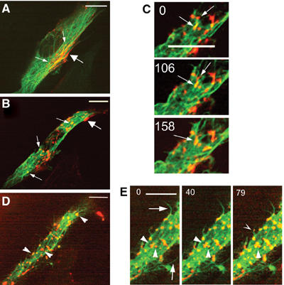
Figure 7.

Myosin organization in cytochalasin D-treated myotubes. Myotubes differentiated for 36 h and expressing GFP-tubulin and Dsred-myosin were imaged at 13-s intervals by spinning disk confocal microscopy for 48 min. Isolated images of movie 6_1 show a myotube displaying myosin localized in the cytoplasm (A, small arrows) and under the plasma membrane (A, large arrow). At 10 min after cytochalasin D addition (B, movie 6_2) myosin fibers disappeared from the plasma membrane (B, large arrow), while myosin structures remained on MTs (B, small arrows) and fused as exemplified in (C) (arrows). At 45 min after drug addition, myotubes dispayed large myosin aggregates (D, arrowheads). Formation of these structures was followed (E, arrowheads), as well as myosin structures (E, hollow arrowhead) localized on growing MT (E, arrows). Time 0 indicates the initial location of selected myosin structures. Time is in seconds, bar=10 μm.
