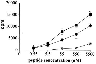
FIG. 3.
In vivo induction of CD4+-T-cell responses by MalE/ACT proteins. C57BL/6 mice were intravenously injected with 50 μg of MalE/ACT proteins bearing the MalE epitope at site 108 (•) or 336 (▪). Mice were injected with mock AC toxoid (asterisks) as negative control. One week later, the mice were sacrificed and the splenocytes were in vitro stimulated with various concentrations of the MalE peptide. After 72 h of culture, [3H]thymidine (50 μCi/well) was added and cells were harvested 6 h later with an automated cell harvester. Incorporated thymidine was detected by scintillation counting. Results show the antigen-specific proliferation obtained for one representative mouse out of four tested in two independent experiments. Each point was done in triplicate and standard deviations are indicated by the bars (n = 3). Results are expressed in changes in counts per minute of incorporated [3H]thymidine (counts per minute in the presence of peptide − counts per minute in the absence of peptide).

