
Figure 2.
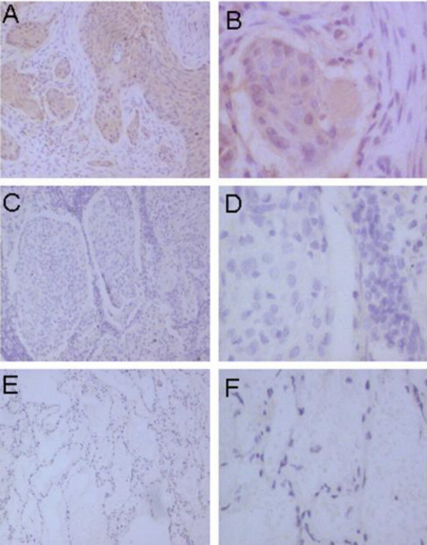
Immunohistochemical staining for PlGF in squamous cell carcinoma of the lung. A and B showed strong diffuse cytoplasmic staining of PlGF in squamous carcinoma of the lung. C and D showed negative staining of PlGF in squamous carcinoma of lung. E and F showed the negative staining status of PlGF in alveolar. (Original magnification ×400)