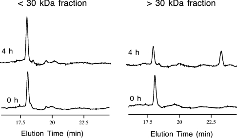
Figure 3. Isomerization of DLP-4 to DLP-2.
RP-HPLC traces of incubation mixtures obtained at the start (0 h) and after 4 h of incubation at 33 °C. The two venom-gland fractions were separated by centrifugal ultrafiltration with a 30 kDa nominal molecular mass cut-off filter. The incubation mixture contained the extract fraction, PBS, DLP-4 and EDTA in the amounts described in the Materials and methods section. The small increase in the DLP-4 HPLC peak after 4 h in the <30 kDa fraction was caused by inaccuracies in injection volume.

