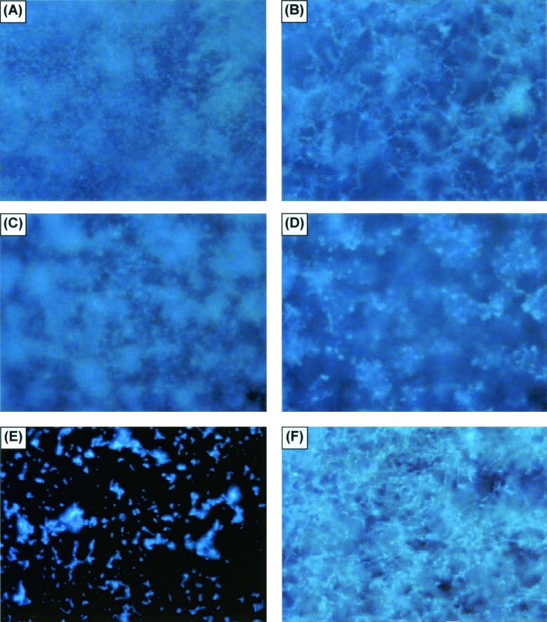
FIG. 6.
FM examination of fungal biofilms formed by different Candida species. (A and B) FM examination of the basal and upper layers of a C. albicans biofilm, respectively. The biofilm was stained with Calcofluor White and viewed at ×10. (C and D) Similar views of C. parapsilosis, although the view of the upper layer is at a lower altitude than that in panel B, due to the limited thickness of the matrix. (E and F) Views of C. glabrata and C. tropicalis, respectively. Other samples of C. tropicalis and C. parapsilosis had the same appearance as that in panel E.

