FIG. 7.
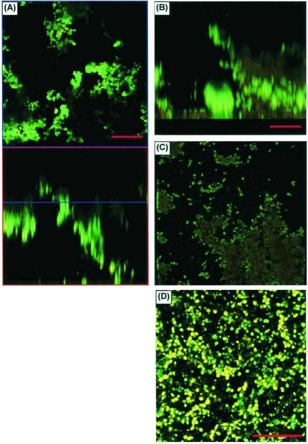
CSLM characterization of C. parapsilosis biofilms. (A) Horizontal (top) and vertical (bottom) slice reconstructions of a C. parapsilosis biofilm examined by CSLM with CAAF and FUN-1. Images were obtained with a ×20 water immersion objective. Green haze is an out-of-focus artifact, not extracellular matrix. (B) Image reconstructed by using transparent projection to show the lateral aspect, which is ≈117 μm deep. (C) Top-down view of the same image stack. (D) View of a different C. parapsilosis isolate, which failed to form appreciable matrix.

