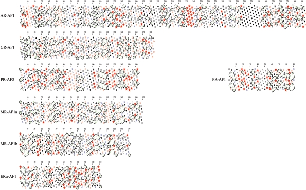
Figure 4. HCA of the SHR-transactivation domains (AF1 and AF3).
HCA is based on pattern recognition and allows for the identification of potentially shared structural folds. The amino acid sequence of the individual transactivation regions has been duplicated and represented on a two-dimensional α-helix [145,146]. Hydrophobic residues (in green) are enclosed by black lines, and proline (red stars), glycine (filled black diamonds), serine ( ), threonine (□) and cysteine (C) residues are highlighted.

