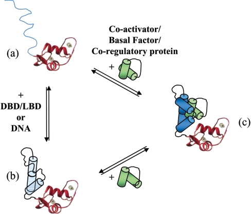
Figure 5. Model illustrating the putative conformers adopted by SHR-NTD.
(a–c) The NTD is represented by line or helical structures (shown in blue) linked to the folded globular structure of the DBD. The binding of the target protein is shown in green. The NTD has regions of significant intrinsic disorder (a), which may be stabilized by other receptor domains, or on binding DNA (b). The NTD can be thought of as a number of conformers with a flexible structure. The binding of a target protein (in green) induces and/or stabilizes the conformation of the SHR-NTD (c).

