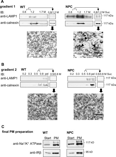
Figure 3. Isolation of PM fractions from WT and NPC mouse livers.
Plasma membranes were isolated by sequential density-gradient centrifugation as detailed in the Experimental section. (A, B) Depletion of contaminating membranes in the preparation. The distribution of lysosomal and ER marker proteins LAMP1 and calnexin respectively in the gradient fractions and interphases was analysed by Western blotting. Interphases represent the membrane fractions collected from gradient 1 and subjected to gradient 2 (0.8/1.2 M) (A), and the final plasma-membrane fraction was collected from gradient 2 (0.5/0.8 M) (B). Electron micrographs of the interphases are also shown. Arrowheads indicate storage organelles. Suc, sucrose; pel, pellet. Equal volumes of the gradient fractions were used for Western-blot analysis. (C) Enrichment of plasma-membrane proteins in the preparation. Na+/K+-ATPase and IR were immunoblotted from the starting material (‘Start’; crude liver lysate subjected to gradient 1) and from the final plasma-membrane fraction (PM). To be able to compare the band intensities in the same blot, 40 μg of protein was used for Start and 10 μg for plasma membrane.

