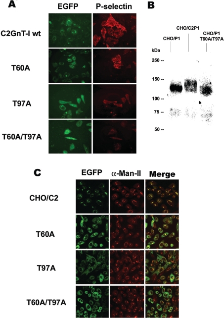
Figure 3. Comparison of P-selectin-binding and subcellular distribution between C2GnT-I and its N-glycosylation-deficient variants.
(A) CHO/F7P1 cells were transiently transfected by DNAs coding for the wt C2GnT-I or its glycomutants (EGFP) and incubated with P-selectin–IgM chimaeras. The binding was then revealed by fluorescence microscopy using an RITC (rhodamine isothiocyanate)-conjugated anti-human IgM secondary antibody. Gain and exposure time were kept constant between images. (B) Western-blot analysis of PSGL-1 synthesized in CHO cells in the absence of C2GnT-I (CHO/P1) or in the presence of the enzyme (CHO/C2P1) or the double mutant T60A/T97A (CHO/P1 T60A/T97A). Proteins were separated by SDS/PAGE followed by immunoblotting using the anti-PSGL-1 mAb PL1. (C) Confocal microscopy images of EGFP-conjugated proteins and α-Man-II immunostaining of cells expressing the wt (CHO/C2) or the mutants T60A, T97A and T60A/T97A. The images shown are the Z sections at 5 μm from the top of the cells. The results are representative of two independent experiments.

